I. Introduction
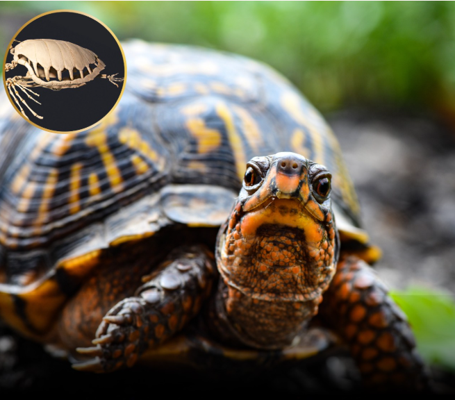
For many, a turtle shell conjures images of a hard, protective helmet; a rigid, lifeless covering that functions merely as armor. But nothing could be farther from the truth. A turtle’s shell is an intricate, living structure—part bone, part keratin, part nervous system—that plays an indispensable role in respiration, skeletal integrity, sensory perception, and overall homeostasis.
This article delves into the remarkable complexity of the turtle shell. We’ll explore anatomical details—how fused ribs and spine form the carapace and plastron—and how nerve endings embedded in that structure mean the shell is capable of feeling. We’ll discuss how damage constitutes injury comparable to a broken limb, how cracks and wear demand veterinary care, and why responsible handlers must treat the shell as an integral part of the living animal.
II. Shell Anatomy & Development: Fused Ribs, Spine, and Keratin Covering
1. Bony Framework: Carapace and Plastron
At the core of the turtle shell are the carapace (upper back) and plastron (ventral belly). These aren’t external attachments—they result from fusion and ossification of ribs, vertebrae, and certain dermal bones. In turtle embryos, ribs grow outward and fuse laterally, integrating with the neural and costal bones. The vertebral column becomes embedded within the carapace, embodying the spine.
On a histological level, the bones are living tissue—vascularized, cellular, constantly remodeling via osteoblast and osteoclast activity—much like other bones in the skeleton. This essential osteology is responsible not just for support but also for growth, repair, and maintaining calcium homeostasis throughout life.
2. Keratin Layer: Scutes and Epidermal Covering
Overlaying the bony scaffold are scutes—keratinized plates reminiscent of fingernails. These are composed of alpha‑keratin (similar to hair and horns). They protect the more fragile bone, reduce wear, and give the shell its often-patterned appearance.
Every scute receives sustenance from the living tissue beneath. The interface between scute and bone is dynamic: scutes grow, wear down, and are replaced. In species like the red-eared slider, growing scutes intermittently peel off to reveal new ones beneath, akin to molting.
III. Nervous Integration: How the Shell Really Feels
Contrary to popular belief, the shell isn’t anesthesia‑like in sensation. Many nerve endings penetrate the periosteal layer and run through the bone towards the surrounding tissue. These nociceptors and mechanoreceptors detect pressure, vibration, and damage.
In controlled studies of rehabilitation and veterinary care, turtles react to shell stimulation—gentle pressure elicits pulling, withdrawal of limbs, or vocalization (in some species). In rescue settings, mild scratching along intact scutes is often reported to produce a quiescent, contented response. That suggests sensory integration that parallels other dermal structures.
When the shell is fractured or cracked, nerve-rich bone tissue is exposed. The turtle may exhibit withdrawal, altered posture, refusal to move, or signs of stress—demonstrating that shell sensation is central to their physiology.
IV. Damage & Pathology: Shell Trauma as Serious Injury
1. Types of Shell Trauma
Shell injuries range from minor chips to catastrophic full-thickness fractures:
- Superficial scute abrasions: Often cosmetic—but can expose bone layer and risk infection.
- Cracks or splits: Partial or full bone fractures exposing internal tissues.
- Punctures and bites: Trauma from predators, accidental crossbite, or domestic pet encounters.
- Burns or chemical damage: Exposure to hot surfaces, ultraviolet over‑irradiation (UV‑C sterilizers), or cleaning agents.
Each requires medical evaluation. Veterinarians specializing in herpetology use radiographs (X‑rays), CT scanning, and sometimes ultrasound to assess depth, displacement, and involvement of internal organs (e.g. lungs slightly beneath the carapace).
2. Healing & Complications
Bone healing in turtles involves callus formation, osteoblast recruitment, and internal remodeling. However, healing is slower than in mammals—sometimes taking months. Persistent moisture or suboptimal thermal conditions can delay repair. If scute coverage does not regenerate properly, exposed bone becomes susceptible to osteomyelitis (bone infection).
To prevent infection and promote healing, standard care includes:
- Cleaning with antiseptics suitable for reptiles (e.g. dilute chlorhexidine or povidone-iodine, used carefully).
- Application of marine-grade epoxy or veterinary-grade resins to stabilize cracks.
- Use of antibiotic therapy—oral or injectable (e.g. ceftazidime or enrofloxacin) under veterinary dosage guidance.
- Supportive husbandry: thermoregulation at optimal basking temperature (usually 28‑32 °C for many species), proper UV‑B lighting for vitamin D₃ synthesis and calcium metabolism, and balanced diet rich in calcium and phosphorus (e.g. calcium‑fiber ratios, supplementation with calcium carbonate or ground cuttlebone).
3. Comparative Severity: Shell Fracture vs. Broken Bone
In turtles, shell fracture is treated with similar gravity as a compound fracture of the limb in mammals:
- Both breach protective tissue, exposing internal structures to contamination.
- Internal hemorrhage may occur if the fracture extends near the coelomic cavity.
- Pain is real and must be managed, though veterinarians may be cautious with analgesics (some opioids or NSAIDs carry risk).
Behavioral changes—lethargy, anorexia, reluctance to bask—are hallmarks of serious shell injury in a turtle.
V. Shell as Vital Physiology: Beyond Protection
1. Respiration & Cardiovascular Roles
While turtles breathe via lungs, the rigid shell restricts rib‑cage movement. Instead, respiration is accomplished through muscular action—the contracting and relaxing of muscles between the pectoral and pelvic girdles, such as transversalis, tricostalis inferior, and oblique abdominal muscles—which shift viscera inside the shell.
In turtles like the spotted turtle and Eastern box turtle, the flexibility between carapace and plastron (via the hinge joint) aids in exhalation/inhalation. The shell’s morphology facilitates negative pressure ventilation rather than the rib-based expansion seen in mammals.
Additionally, the shell serves as a surface for vascular exchange. Blood vessels close to the keratin layer can function in thermoregulation—distributing heat absorbed from basking or releasing heat during cooling.
2. Mineral Storage & Calcium Homeostasis
The shell is a calcium reservoir. During periods of egg‑laying (oviposition) or when dietary calcium is insufficient, the turtle mobilizes minerals from the shell via osteoclastic resorption and systemic circulation. This is crucial to avoid metabolic bone disease (MBD).
Veterinary monitoring of calcium‑phosphorus levels, along with assessment of shell density via radiographs, helps track bone mineralization. In rehabilitation centers, veterinarians may administer calcitriol (active vitamin D₃) or calcium gluconate titration to support shell and skeletal health.
VI. Shell Sensitivity & Handling Considerations
1. Behavioral Signs of Shell Discomfort
Turtles have varied responses to shell contact:
- Some individuals enjoy gentle stroking along the scutes, remaining still or holding their limbs open—similar to purring in cats.
- Others recoil quickly, tuck limbs, or retract head when the scutes are scratched.
- Overly aggressive handling may cause stress responses such as hissing, bead‑rolling eyes, or biting.
Handlers—zoologists, veterinarians, rehabilitators—are advised to watch for each turtle’s individual threshold.
2. Handling Protocols for Welfare
To minimize risk:
- Always support the shell evenly, avoiding pressure points.
- Use two-handed lifts, one under carapace and one under plastron.
- Never pick by the scutes or allow the turtle to dangle unsupported.
- When examining for minor scute damage, provide warming to enhance circulation and make tissues less brittle.
- For cleaning, use lukewarm water and soft cloth, minimizing abrasive mechanical stress.
VII. Evolutionary Origins & Comparative Biology
1. Fossil Record & Morphogenetic Adaptations
The turtle shell is a unique evolutionary innovation dating back roughly 220 million years (Late Triassic). Transitional fossils like Proganochelys show early development: partial ossifications and limited spinal integration. Over millennia, turtles evolved a unique axial skeleton where ribs expand laterally and the lungs become dorsal to internal organs.
The integration of dermal and endoskeletal bones (neural, costal, peripheral) into a unified shell is a marvel of developmental morphogenesis. Genes like Sonic hedgehog (Shh), Msx1/2, and Bmp family members orchestrate rib outgrowth and keratinization.
2. Diversity of Shell Morphologies
Across species, shell shape, scute arrangement, hinge flexibility, and coloration vary dramatically:
- Softshell turtles (family Trionychidae)—leather-like skin instead of keratin scutes.
- Tortoises—domed, thick carapace with heavy bone mass for terrestrial defense.
- Sea turtles—streamlined carapace with reduced scutes, optimized for aquatic hydrodynamics.
- Box turtles (genus Terrapene)—plastral hinge allowing full closure, offering defense and thermoregulation.
Each evolutionary path aligns shell form with habitat, predator pressure, reproductive strategy, and metabolic investment.
VIII. Shell Regeneration & Rehabilitation Techniques
1. Clinical Assessment & Radiographic Diagnostics
Veterinarians rely on:
- Digital radiography (X‑ray) for evaluating bone continuity and shell density.
- CT scans for 3D reconstruction of fractures.
- Blood panels measuring calcium, phosphorus, uric acid, and vitamin D metabolites.
- Microbiological cultures from exposed bone to guide antibiotic therapy.
2. Surgical Intervention & External Repair
For partial-thickness fractures:
- Epoxy or fiberglass patching—stabilizes edges, prevents ingress of pathogens.
- Wire cerclage or surgical pins—in deeper or displaced bony fractures, with careful avoidance of pulmonary cavity.
Click page 2 for more


